




Dubai knee assessment
Knee Pain When Bending or Squatting: Find the Pattern Before You Treat It
Pain in a squat, on stairs or during deep knee bend can come from the kneecap, tendon, meniscus, cartilage or an injury-related instability pattern. The useful question is not simply “what injection or scan do I need?” It is: what structure and loading pattern best explain this knee?
- Location mattersFront, joint-line, below kneecap or instability-related pain can point in different directions.
- Symptoms guide urgencyLocking, rapid swelling, inability to bear weight, or a hot joint deserve prompt assessment.
- Imaging is targetedX-ray, ultrasound or MRI should answer a clinical question—not replace the examination.
The direct answer
Knee pain with bending is common. The correct treatment depends on the pattern, not the movement alone.
A painful squat can reflect patellofemoral overload, a patellar tendon problem, a meniscus-related symptom pattern, cartilage or osteoarthritis change, an acute ligament injury, or a less common inflammatory or bony condition. Pain location, onset, swelling, locking, instability and your training history help narrow the possibilities.
Why a deep bend can hurt
Squatting increases demand across more than one knee structure at the same time.
As the knee bends, the patellofemoral joint, tibiofemoral joint, menisci, cartilage, tendons and muscles all contribute to controlled movement. A squat may expose a mismatch between tissue capacity and training demand, but it can also reveal a structural injury after a twist or impact.
That is why “squat pain” is not enough to label the problem. A precise plan starts by matching the symptom pattern to the clinical examination.
Knee pain map
Seven clinical patterns worth separating
These patterns overlap. They are not a self-diagnosis tool, but they explain why the same squat can be painful for different reasons in different people.
Front-of-knee pain
Pain around or behind the kneecap with stairs, running, long sitting, squats or getting up from a chair can fit a patellofemoral pain pattern.
Patellofemoral assessmentPinpoint pain below the kneecap
Focal pain at the patellar tendon—often during jumping, sprinting, hills or heavy gym work—can fit a tendon-load problem rather than a joint problem.
Tendon injury careJoint-line pain after a twist
Inner- or outer-joint-line pain, delayed swelling, catching or pain with deep bend and twisting may suggest a meniscus-related problem.
Meniscus and cartilage careDeep ache, stiffness or recurrent swelling
Cartilage sensitivity or osteoarthritis may present with activity-related ache, stiffness after rest and swelling that returns after heavier loading.
Cartilage-focused reviewGiving way after a pivot or landing
Shifting, instability or loss of confidence after trauma should raise concern for ligament injury or a combined internal-knee injury pattern.
ACL and ligament careKneecap shift or dislocation history
A feeling that the kneecap slips, shifts or has dislocated can be different from ordinary anterior knee pain and may need a specific instability assessment.
Kneecap instability assessmentUnusual, severe or worsening symptoms
Rapid progression, night pain, fever, redness, marked warmth, focal bone pain or pain after a significant trauma needs broader assessment—not a “push through it” plan.
Read urgent signsRed flags
Do not keep testing a painful knee when the symptom pattern suggests a potentially urgent problem.
These signs do not diagnose the cause alone, but they justify prompt clinical assessment. A knee that is mechanically blocked or a hot, acutely swollen joint should not be managed as routine gym soreness.
Diagnosis first
A scan can be helpful. It is rarely the diagnosis on its own.
The purpose of assessment is to determine whether a painful bend is primarily a load-management problem, an intra-articular problem, an instability problem or an urgent condition requiring a different pathway.
- 01
Understand the onset and trigger
Gradual training-related pain is different from a sudden twist, pop, fall, direct blow or first-time kneecap dislocation.
- 02
Examine the knee in motion
We assess swelling, range, joint-line tenderness, stability, tendon sensitivity, kneecap mechanics, strength and functional movement.
- 03
Use imaging to answer a question
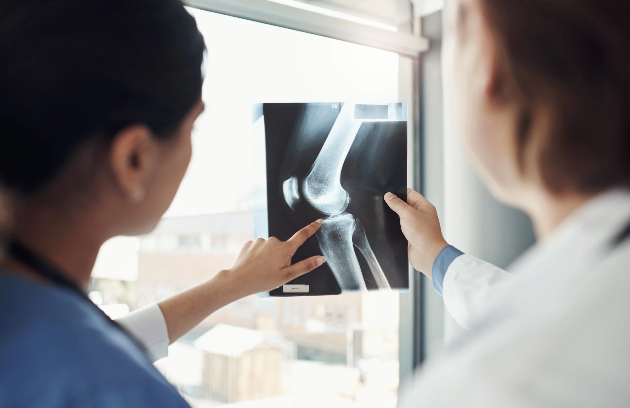
X-ray helps with bone, alignment and arthritis assessment. Ultrasound can help with superficial tendon or bursal questions. MRI is useful for meniscus, cartilage, ligaments, bone-marrow and deeper soft-tissue questions when it will change management.
- 04
Match the plan to the target
Rehabilitation, activity modification, bracing, medication, injection or surgery are tools. The correct choice depends on the clinically relevant problem.

What you can do now
Protect the irritated task without making the whole knee inactive.
These are general steps for non-urgent situations. They do not replace assessment after significant trauma, true locking, marked swelling or a red-flag symptom pattern.
Modify depth and load
Temporarily reduce the squat depth, load, jumping volume, kneeling or pivoting task that clearly provokes symptoms. Avoid repeatedly testing the exact painful range.
Keep appropriate movement
Comfortable walking, cycling, mobility or lower-load strengthening may be useful when they do not cause meaningful worsening, swelling or instability.
Watch the response
Sharp pain, an enlarging effusion, loss of movement, new locking, giving way or symptoms that are progressively worsening are reasons to stop and reassess.
Do not self-prescribe a procedure
A brace, medication or injection may help selected cases, but should be linked to diagnosis, medical history and a rehabilitation plan—not simply to a painful squat.
Treatment pathways
The right treatment is determined by the diagnosis—not by the word “squat.”
Most non-urgent knee pain does not start with surgery. But rehabilitation should not delay treatment when a true mechanical block, unstable injury or another clear surgical target is present.
Injections can be considered in selected diagnoses. They should not replace a clear explanation of the knee problem or a rehabilitation pathway.
FAQ
Common questions about pain with bending and squatting
These answers are educational. The same symptom can have different causes depending on age, injury story, location of pain, swelling, examination and imaging when needed.
Is knee pain when squatting always a meniscus tear?
No. Pain with a squat can come from the kneecap, patellar tendon, meniscus, cartilage, arthritis, instability or loading mechanics. A meniscus problem becomes more plausible with joint-line pain after a twist, swelling, catching or true locking, but symptoms alone do not confirm it.
Should I stop squatting completely?
Not always. In many non-urgent overload patterns, a temporary change in depth, load, tempo or training volume is more useful than total rest. Stop and seek assessment if there is sharp pain, meaningful swelling, locking, instability or worsening symptoms.
Why does my knee hurt going downstairs but not on flat ground?
Descending stairs increases demand through the patellofemoral joint and requires controlled quadriceps work. That pattern is often seen with front-of-knee pain, but tendon, cartilage or other conditions can also contribute.
When is an MRI useful for knee pain when bending?
MRI is useful when a meniscus, cartilage, ligament, bone-marrow or deeper soft-tissue question is suspected and the answer is likely to change the plan. It is not automatically necessary for every gradual knee pain presentation.
Can PRP fix knee pain when bending?
PRP may be discussed in selected diagnoses, but it is not a universal treatment for squat pain. It does not correct a locked knee, restore a clinically unstable complete ligament or replace a mechanical treatment when surgery is indicated.
When should I get urgent help?
Seek prompt medical assessment for a knee that is truly locked, a major swelling after trauma, inability to bear weight, a deformity, a hot red swollen joint with fever, or significant instability after injury.
Clear diagnosis. Appropriate next step.
Persistent squat pain deserves a plan that matches your knee, your activity and your goals.
Dr. Tomislav Cerovecki provides knee assessment, MRI review, rehabilitation planning, injection discussion when appropriate and surgery only when there is a clear indication at Tadawi Hospital, Al Garhoud, Dubai.