




Knee imaging · patient guide
MRI for Knee Pain in Dubai: When Do You Really Need It?
MRI can be very useful when knee pain may involve a meniscus tear, ACL injury, cartilage damage, bone bruise, hidden injury, persistent swelling or loss of function. But MRI is not automatically the first step — and a scan should always be interpreted together with symptoms, physical examination and your individual goals.
- MRI shows important internal soft-tissue structures in detail.
- X-rays may be more useful first for many acute injuries, fracture concerns or arthritis assessment.
- The best scan is the one that changes the next treatment decision.
The essential answer
MRI is most valuable when it can change the treatment plan.
MRI can provide detailed information about internal knee structures. However, a scan is only one part of the assessment. The most useful decision comes from understanding how the injury happened, where the pain is, whether the knee swells or locks, how stable it feels, and what you need to return to in daily life, work or sport.
Symptoms first
Swelling, true locking, instability, reduced movement and loss of function matter.
Examination plus imaging
MRI helps identify structures. Examination shows how the knee behaves in real life.
Decision-focused care
The goal is not simply to obtain a scan, but to choose the right next step.
Understanding the scan
What Can an MRI Show in Knee Pain?
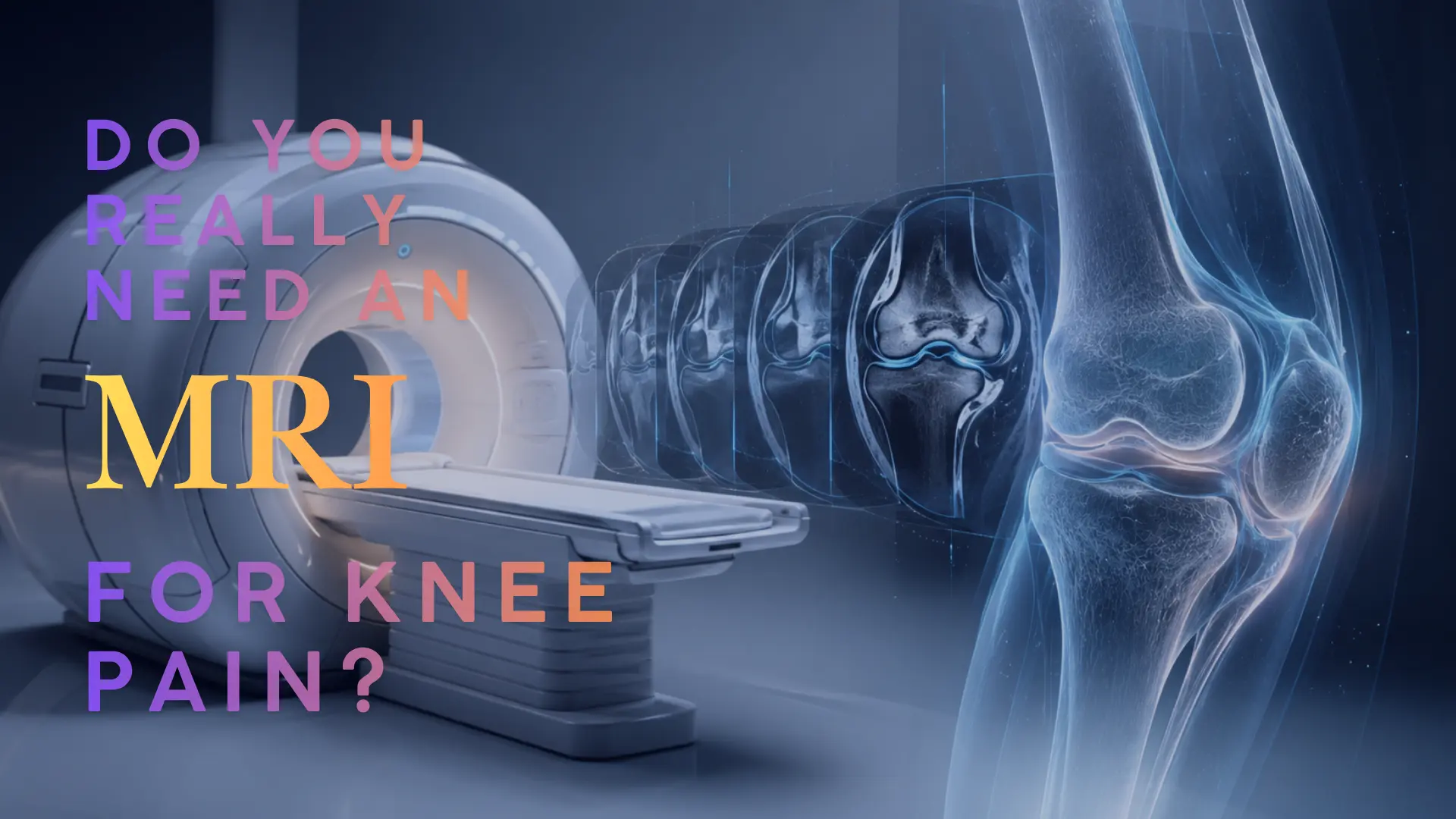
MRI uses a strong magnetic field and radio waves to create detailed images of structures inside the knee. It is especially useful when the clinical question involves soft tissue, cartilage, bone marrow or deeper joint structures that cannot be assessed fully with an X-ray alone.
A knee MRI can help assess the meniscus, ACL and other ligaments, joint cartilage, bone bruising, bone-marrow changes, joint fluid, selected tendon problems and some hidden injuries. The important question remains whether the MRI finding explains your symptoms and changes the appropriate treatment plan.
Meniscus tears
MRI can help identify the location and pattern of a meniscus tear. A tear on the scan does not automatically mean surgery; symptoms, locking, cartilage health, knee function and whether repair is realistic all matter.
Explore meniscus and cartilage careACL and ligament injuries
MRI is useful for assessing ACL and other major ligament injuries, particularly after a pivot, landing, sudden stop or twisting injury. It can also identify associated meniscus, cartilage and bone-bruising findings.
Explore ACL and ligament careCartilage and kneecap conditions
MRI can show focal cartilage defects, cartilage thinning and selected kneecap findings. These changes must be interpreted carefully because pain can also relate to overload, muscle weakness, alignment or kneecap tracking.
Explore kneecap and cartilage conditionsBone bruising and hidden injury
MRI can help detect bone bruising, bone-marrow changes and selected occult injuries when symptoms remain unexplained after initial assessment. It can also show joint fluid and selected inflammatory changes.
Explore fractures and sports trauma careVideo explanation
When does an MRI help in knee pain?
Dr. Tomislav explains what MRI can show, what it cannot decide alone, and why the report must always be matched to symptoms and examination.
- Meniscus, ACL and cartilage findings
- When a scan may change treatment
- Why the report is not the final diagnosis
Decision points
When Is MRI for Knee Pain Usually Helpful?
Not every painful knee needs MRI immediately. MRI becomes more useful when the result may clarify an important structural concern or guide a different treatment pathway.
After a significant twisting or sports injury
MRI may be useful after a meaningful twist, pivot, fall, landing or direct impact, especially when there is early swelling, instability, loss of movement or concern for ligament, meniscus or cartilage injury.
When the knee is truly locking or cannot fully move
A knee that repeatedly gets stuck, cannot fully straighten or has a clear mechanical block needs prompt clinical assessment. MRI may help identify a displaced meniscus tear, loose body or another internal cause.
When swelling, instability or loss of function persists
Recurrent swelling, repeated giving way, reduced confidence or ongoing limits with walking, stairs, squatting or sport may justify further imaging after an appropriate examination.
When a suitable first-line plan has not helped
When symptoms continue despite a structured plan of load modification, rehabilitation, strengthening and clinical review, MRI may help clarify whether there is a deeper structural problem.
When a procedure or surgery is being considered
MRI can support treatment planning by showing the meniscus, cartilage, ligaments and associated structures in greater detail. It supports planning; it is not the only reason to recommend surgery.
Choosing the right test
X-ray, MRI or Ultrasound: Which Test Answers Which Question?
These tests are not competitors. Each test provides different information, and the correct choice depends on the symptoms, examination and clinical question.
Bone, alignment and arthritis
Often useful early for suspected fracture, arthritis, joint-space narrowing, bone alignment and selected kneecap or bony conditions.
Internal soft tissues
Most useful for meniscus, ligaments, cartilage, bone bruising, bone-marrow changes and deeper structures when the result can guide management.
Selected superficial structures
Useful for selected tendons, fluid collections, dynamic assessment and image-guided procedures when clinically appropriate.
Clinical interpretation
An MRI Report Is Important — But It Is Not the Full Diagnosis
MRI describes what is visible inside the knee. The orthopaedic diagnosis connects those findings to your symptoms, physical examination, injury mechanism, functional limits and goals.
A good knee diagnosis combines:
Two patients can have similar MRI reports but need different treatment. For example, a fresh traumatic meniscus tear with true locking in an active athlete is not the same clinical situation as a small degenerative tear with early arthritis symptoms.
Can MRI decide whether surgery is needed?
No. A meniscus tear, cartilage change or ligament injury on MRI does not automatically mean surgery. Surgery is considered when the full clinical picture supports it — such as true mechanical locking, recurrent instability, a repairable acute injury, significant functional limitation or symptoms that have not improved with appropriate non-surgical care.
Urgent assessment
Do not wait for a routine MRI appointment when urgent warning signs are present.
Seek urgent medical assessment after an injury if there is obvious deformity, inability to bear weight, severe or rapidly increasing swelling, a cold or numb foot, a hot red knee with fever, severe pain after trauma, or a knee that is truly locked and cannot move normally.
Patient questions
Common Questions About MRI for Knee Pain
Do I need an MRI for every knee pain?
No. Many knee problems can be assessed clinically first. MRI is usually considered when internal injury is suspected, symptoms persist, there is swelling, instability, true locking, loss of function, or the result may change the treatment plan.
Can MRI show a meniscus tear?
Yes. MRI can often identify a meniscus tear and help show its location and pattern. But a meniscus tear on MRI does not automatically mean surgery; symptoms, age, cartilage health, function and tear pattern all matter.
Can MRI show an ACL injury?
Yes. MRI is useful for assessing ACL injury and associated findings such as bone bruising, meniscus tears, cartilage injury or injury to other stabilising structures around the knee.
Is MRI better than X-ray for knee pain?
They answer different questions. X-rays are useful for bone alignment, fractures and arthritis. MRI is better for soft tissues such as meniscus, ligaments, cartilage, selected tendons and bone bruising. The right test depends on the clinical situation.
Should I bring my MRI images and report to my consultation?
Yes. Bring the written report and, whenever possible, the original images or access link. This allows the scan to be reviewed with your symptoms, examination findings and treatment goals.
Orthopaedic consultation in Dubai
Not sure whether MRI is the right next step for your knee pain?
Dr. Tomislav can assess your symptoms, examine your knee, review previous X-rays or MRI scans, and explain the most appropriate next step for your individual condition.
- Doctor
- Dr. Tomislav Cerovecki
- Clinic
- Tadawi Specialty Hospital, Al Garhoud, Dubai
- Useful for
- Knee pain, MRI review, sports injuries, meniscus, ACL and cartilage concerns.
Medical note: This article is for general educational purposes only. It does not replace a personal consultation, diagnosis or treatment plan. MRI findings, symptoms and treatment options vary between individuals.