






Patient guide · Complex knee ligament injuries
PCL, MCL, LCL and Multi-Ligament Knee Injuries: Symptoms, MRI and Treatment in Dubai
A knee with one injured ligament may need protection and rehabilitation. A knee with several major stabilisers damaged may require urgent assessment and a very different treatment plan. The important question is not simply how many tears appear on MRI, but how the whole knee behaves.
- Written by
- Dr. Tomislav Cerovecki
- Clinical focus
- Sports traumatology, knee surgery and complex instability
- Clinic
- Tadawi Specialty Hospital, Al Garhoud, Dubai

The essential answer
A multi-ligament knee injury is not one standard diagnosis and does not automatically mean one standard operation.
It generally means that at least two major ligament complexes are injured. Treatment depends on which structures are damaged, whether the knee is mechanically unstable, whether circulation or nerves are affected, and which tissues can heal with bracing and rehabilitation.
Start with the whole injury
One MRI can contain several findings, but they do not all carry the same weight.
When an MRI lists a PCL tear, MCL injury, LCL damage or a posterolateral corner injury, the terminology can feel overwhelming. It is natural to assume that every listed tear needs surgery. That is not how these injuries are assessed.
An isolated MCL injury may heal successfully with a hinged brace and structured rehabilitation. A lower-grade isolated PCL injury may also recover without an operation. By contrast, a complete PCL tear combined with major posterolateral corner instability creates a different mechanical problem and is more likely to require reconstruction.
The clinical task is to understand which structures are truly unstable, which structures still have healing potential, and whether the knee can safely regain function without leaving persistent abnormal movement.
The MRI report is a map, not the final treatment decision. The injury mechanism, examination, stress testing, circulation, nerve function and the patient’s activity goals all matter.
Four major stability systems
What the PCL, MCL, LCL and posterolateral corner do.
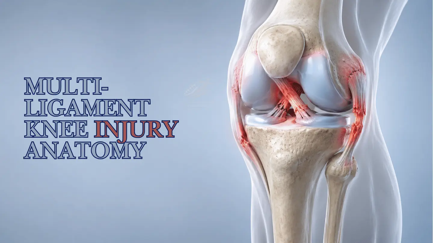
The ligaments work together to control backward movement, sideways opening and rotation. A combined injury can therefore feel different from an isolated tear.
Posterior cruciate ligament
The PCL sits inside the knee behind the ACL. Its principal role is to limit excessive backward movement of the shinbone relative to the thighbone.
A classic mechanism is a direct impact to the front of a bent knee, such as a dashboard injury, a fall onto the flexed knee or contact during sport.
Medial collateral ligament
The MCL lies on the inner side of the knee and resists excessive opening of the medial joint when a force pushes the knee inward.
It is commonly injured by a blow to the outer knee, twisting, skiing injuries, contact sport or an awkward inward collapse during landing.
Lateral collateral ligament
The LCL lies on the outer side of the knee and helps resist forces that push the knee outward.
A significant apparent LCL injury should prompt assessment of the wider posterolateral corner, because the LCL rarely acts alone in severe outer-knee instability.
Posterolateral corner
The PLC is a group of stabilising structures at the back and outer side of the knee. It controls outward angulation and external rotational instability.
Missing a major PLC injury can leave ongoing instability and place excessive stress on an ACL or PCL reconstruction.
A broad clinical category
What is a multi-ligament knee injury?
It generally describes injury to at least two major ligament complexes. The term includes a wide spectrum, from combined sporting injuries to severe trauma.
Possible combinations include ACL and MCL, PCL and MCL, PCL and posterolateral corner, ACL and PCL, or injuries involving three or four major ligament regions.
These combinations are not equivalent. A partial ACL injury with a healing MCL sprain is very different from complete disruption of the PCL and posterolateral corner after a road-traffic accident.
The term also does not automatically mean that the knee fully dislocated. Some multi-ligament injuries occur without a documented dislocation. In other cases, the knee may dislocate briefly and move back into position before the patient reaches hospital. This is called a spontaneously reduced dislocation.
A knee that looks aligned can therefore still have significant ligament, vascular or nerve injury. The presence or absence of a documented dislocation should be recorded separately from the ligament pattern.
How the injury occurs
Multi-ligament knee injuries can follow both high-energy trauma and sport.
The direction of force often gives useful clues, but real injuries commonly combine impact, bending and rotation.
Direct impact
A dashboard injury or direct blow to a bent knee can drive the tibia backward and injure the PCL, sometimes together with other structures.
Contact sport
Football, rugby, martial arts and other contact sports can combine valgus, varus, hyperextension and rotational forces.
Twisting or awkward landing
A planted foot, sudden change of direction or poorly controlled landing can injure more than one stabilising structure.
High-energy trauma
Motorcycle injuries, road collisions and falls from height increase concern for fractures, dislocation, vascular injury and nerve damage.
What patients may notice
Symptoms depend on which stability systems have failed.
Possible symptoms include rapid swelling, severe pain, bruising, difficulty bearing weight, reduced movement and a feeling that the knee is shifting or giving way.
Location can provide clues. Inner-knee pain and tenderness are common with MCL injury. Outer-knee symptoms raise concern for the LCL or posterolateral corner. A PCL injury may produce pain or pressure behind the knee and difficulty with stairs, slopes, deceleration or a feeling that the shin is moving backward.
Not every serious injury is dramatically painful. Some PCL injuries are underestimated because there may be no clear pop and the initial symptoms can appear less dramatic than a typical ACL tear.
Do not wait with these signs
When is a knee ligament injury an emergency?
A severe multi-ligament injury can affect the popliteal artery behind the knee or the nerves travelling into the lower leg. A knee that has already moved back into position can still have these complications.
Circulation warning signs
- The foot becomes cold, pale or changes colour
- The pulse at the foot feels weak or absent
- Swelling and pain are rapidly increasing
- The knee was visibly deformed or dislocated
Nerve and trauma warning signs
- Increasing numbness or tingling
- New inability to lift the foot or toes
- An open wound around the injured knee
- Severe trauma with major instability
Seek emergency medical assessment immediately when any of these signs are present. Do not rely on the knee looking aligned or on being able to take a few steps.
Diagnosis is more than one scan
How PCL, MCL, LCL and multi-ligament injuries are assessed.
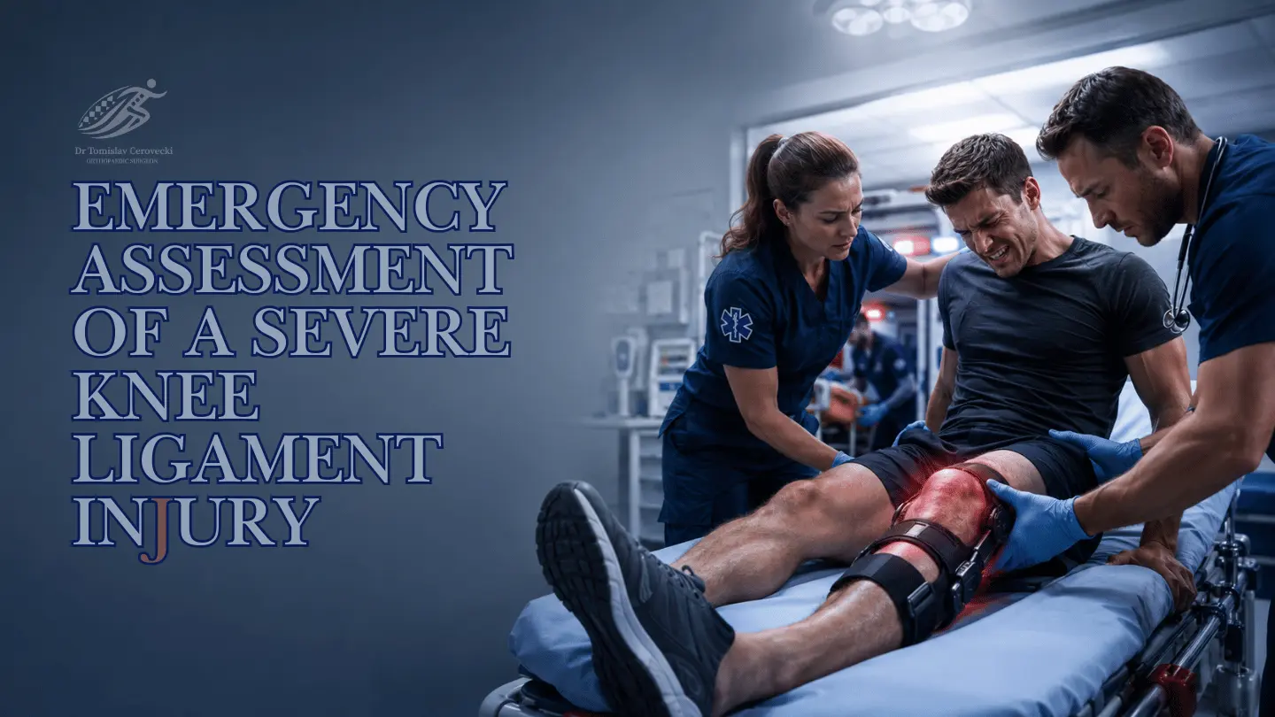
A complete assessment combines the mechanism of injury, repeated clinical examination, imaging and objective measurement of instability when needed.
Injury history
The direction of impact, whether the foot was fixed, whether the knee twisted or hyperextended, whether it looked deformed and how quickly swelling appeared all help define the likely pattern.
Clinical stability tests
The injured knee is compared with the uninjured side. Tests assess forward and backward tibial movement, medial and lateral opening, posterior sag and rotational instability.
Circulation and nerve examination
Pulses, skin temperature, colour, sensation, ankle movement and the ability to lift the foot are checked, particularly after severe trauma or outer-knee injury.
X-rays and stress radiographs
Standard X-rays identify fractures, avulsion injuries and alignment. Stress radiographs can objectively measure how far the knee opens or shifts under a controlled force.
MRI
MRI helps show which ligaments are torn, the tear location and associated meniscus, cartilage, tendon, bone-bruising or corner injuries. It supports—but does not replace—the clinical diagnosis.
Vascular imaging when indicated
CT angiography may be required when the mechanism, examination or injury pattern creates concern for arterial damage, even when the knee is currently aligned.
Why MRI findings and knee instability are not always identical
An MRI can show structural damage, but it does not directly reproduce the forces of walking, turning or sport. A ligament can look abnormal while retaining useful stability, or the knee can demonstrate clinically important abnormal movement that requires a closer review of several structures. Imaging and examination should answer the same clinical question.
Read when MRI helps with knee painTreatment is pattern-specific
Does every PCL, MCL, LCL or multi-ligament injury need surgery?
No. Some isolated injuries heal well with protection and rehabilitation. Complete combined instability is more likely to need surgical reconstruction.
| Injury pattern | Non-surgical treatment may be appropriate when | Surgery may be considered when |
|---|---|---|
| Isolated PCL | The tear is lower grade, the knee remains functionally stable and rehabilitation restores confidence and control. | There is high-grade laxity, persistent instability, bony avulsion, major activity demands or another ligament is injured. |
| Isolated MCL | Most Grade I, Grade II and selected Grade III injuries have a favourable healing pattern with a hinged brace and rehabilitation. | The tissue is displaced or trapped, healing is unlikely, instability remains, or the MCL is part of a more complex injury. |
| LCL / PLC | A stable, low-grade isolated injury may recover with protection, bracing and rehabilitation. | There is complete or rotational instability, poor healing potential, associated cruciate injury or chronic giving way. |
| Multi-ligament injury | Selected lower-grade patterns, medically complex cases or injuries in which some structures can heal may use a combined non-operative plan. | There are complete tears with mechanical instability, young or active patient demands, failed conservative care or associated repairable damage. |
This comparison describes general principles, not a personal treatment recommendation. Tear location, tissue quality, timing, alignment, fractures, skin condition, meniscus damage and neurovascular status can change the plan.
PCL and MCL healing differ
Why some ligaments can be braced while others need reconstruction.
The MCL has a relatively favourable ability to heal because of its location and blood supply. This is why many isolated MCL injuries—even some complete tears—are initially managed with a hinged brace, controlled movement and progressive rehabilitation.
Many isolated lower-grade PCL injuries can also be managed without surgery. Rehabilitation commonly prioritises quadriceps strength because the quadriceps helps control posterior movement of the tibia.
Outer-side injuries require greater caution. A complete LCL or posterolateral corner injury may not heal reliably enough to restore rotational stability, particularly when combined with PCL or ACL damage.
Repair, reconstruction and timing
There is no responsible rule that every injured ligament must be operated on immediately.
A ligament repair uses the patient’s existing tissue and reattaches or sutures it. Reconstruction replaces a damaged ligament with a graft. Repair may be suitable for a recent avulsion with healthy tissue; reconstruction is more commonly considered for midsubstance tears, poor tissue, chronic injury or persistent instability.
Some multi-ligament injuries can be reconstructed in one operation. Other cases need staged treatment because of fractures, severe swelling, restricted movement, skin or soft-tissue damage, vascular treatment or other medical priorities.
During reconstruction, tunnel positions, graft selection and tensioning sequence must be planned as one coordinated system. Failure to recognise a posteromedial or posterolateral injury can overload a cruciate graft and leave the knee unstable.
Restore anatomy
Reconstruction should address the true injured structures rather than only the most obvious MRI label.
Protect movement
Timing balances stable repair with the need to reduce stiffness and recover useful range of motion.
Plan the whole knee
Meniscus, cartilage, alignment, graft tunnels, nerve risk and rehabilitation restrictions must be considered together.
Recovery is part of treatment
Rehabilitation after a multi-ligament injury is longer and more protective than standard ACL rehabilitation.
The exact brace, movement and weight-bearing rules depend on the reconstructed or healing structures. A generic online programme is not enough.
Protect and recover movement
Control swelling, protect repaired structures, restore full extension, begin safe flexion and reactivate the quadriceps according to the surgeon-led protocol.
Rebuild walking and strength
Progress weight-bearing, walking quality, hip and leg strength, balance and basic movement control without provoking instability or excessive swelling.
Restore athletic capacity
Develop strength symmetry, cardiovascular capacity, running tolerance, landing mechanics and controlled change of direction.
Return to work or sport
Use objective testing, sport-specific exposure and psychological readiness rather than relying on the calendar alone.
Return to sport
Nine to twelve months may be a useful general range, but time alone cannot clear the knee.
A safe return may assess swelling, range of motion, ligament stability, quadriceps and hamstring strength, hopping and landing control, running tolerance, change-of-direction ability, sport-specific performance and confidence. A cyclist, construction worker, recreational runner and professional footballer do not place the same demands on the knee.
Why complete assessment matters
The greatest risk is not only missing a tear. It is misunderstanding how the injuries work together.
Complex instability is a system problem. Treating one structure while another major stabiliser remains unrecognised can compromise the result.
A PCL injury can look deceptively quiet
There may be no dramatic pop, and symptoms can be underestimated until stairs, slopes and deceleration expose posterior instability.
An MCL tear can be overtreated
Many MCL injuries can heal with well-managed bracing and rehabilitation. The tear grade alone does not determine surgery.
An LCL label can be incomplete
The wider posterolateral corner and common peroneal nerve must be assessed, particularly with major outer-knee instability.
A cruciate graft can be overloaded
Unrecognised posteromedial or posterolateral instability may place abnormal forces on ACL or PCL reconstruction.
Patient questions
PCL, MCL, LCL and multi-ligament knee injury FAQs
These answers provide general education. They do not replace an individual examination after a significant knee injury.
Can I still walk with a multi-ligament knee injury?
Yes. Some patients can walk, particularly after a dislocated knee has returned to position or when pain is not dramatic. Being able to walk does not rule out serious ligament, vascular or nerve injury. Avoid repeatedly testing an unstable knee and arrange appropriate assessment.
Is MRI enough to diagnose a multi-ligament injury?
No. MRI is highly useful, but the mechanism, clinical stability tests, blood-vessel and nerve examination, X-rays and sometimes stress radiographs or CT angiography are also important.
Can a complete ligament tear heal without surgery?
Some complete MCL tears can heal with appropriate bracing and rehabilitation. Complete PCL, LCL, posterolateral corner and combined injuries behave differently. Tear location, instability and associated damage determine the plan.
Is an LCL tear the same as a posterolateral corner injury?
No. The LCL is one component of the outer stability system. A posterolateral corner injury may involve the LCL together with other structures that control varus and rotational stability.
Does a multi-ligament knee injury always require reconstruction?
No. Some structures may heal with bracing while another is reconstructed, and selected lower-grade patterns may be treated without surgery. Complete combined mechanical instability is more likely to need operative treatment.
How soon should I see a knee specialist?
Seek emergency care immediately for deformity, a cold or pale foot, weak pulses, increasing numbness or new foot weakness. Without these signs, arrange prompt orthopaedic review after a major collision, fall, severe twisting injury or a knee that remains swollen and unstable.
How long does recovery take?
Recovery varies widely. Isolated braced injuries can recover sooner than multi-ligament reconstruction. Return to demanding sport after reconstruction commonly takes around nine to twelve months or longer and should be based on objective milestones rather than time alone.
Clinical basis
Selected medical references
The article reflects current patient-facing orthopaedic principles and international expert consensus. Evidence for complex multi-ligament injuries remains less uniform than for many isolated ligament injuries, so treatment must be individualised.
- Murray IR et al. Multiligament knee injury: expert consensus on nomenclature, diagnosis, treatment and rehabilitation. British Journal of Sports Medicine. 2024.
- Super JT, Chahla J, Geeslin AG, Moatshe G, LaPrade RF. Multiligament Knee Injuries. Journal of Bone and Joint Surgery. 2026.
- American Academy of Orthopaedic Surgeons. Posterior Cruciate Ligament Injuries.
- American Academy of Orthopaedic Surgeons. Collateral Ligament Injuries.
- D’Ambrosi R et al. Return to Sport After Multiligament Knee Injury: A Systematic Review of the Literature. Indian Journal of Orthopaedics. 2024.
Complex knee injury care in Dubai
Get one clear explanation of the complete stability pattern—not a list of disconnected MRI findings.
Dr. Tomislav Cerovecki provides assessment for sports knee injuries, PCL, MCL, LCL and complex ligament instability, including clinical examination, MRI review, individualised treatment planning and return-to-activity guidance.