






MENISCUS & CARTILAGE · PATIENT GUIDE · DUBAI
Meniscus Root Tear: Symptoms, MRI Findings and When Repair Is Considered
A meniscus root tear affects the attachment that helps the meniscus distribute load across the knee. It deserves careful assessment—but the words “root tear” on MRI do not automatically mean surgery.
- A root tear can change normal meniscus mechanics.
- MRI findings should be matched with symptoms, examination, cartilage health and alignment.
- Repair may be considered when preservation is realistic and clinically appropriate.
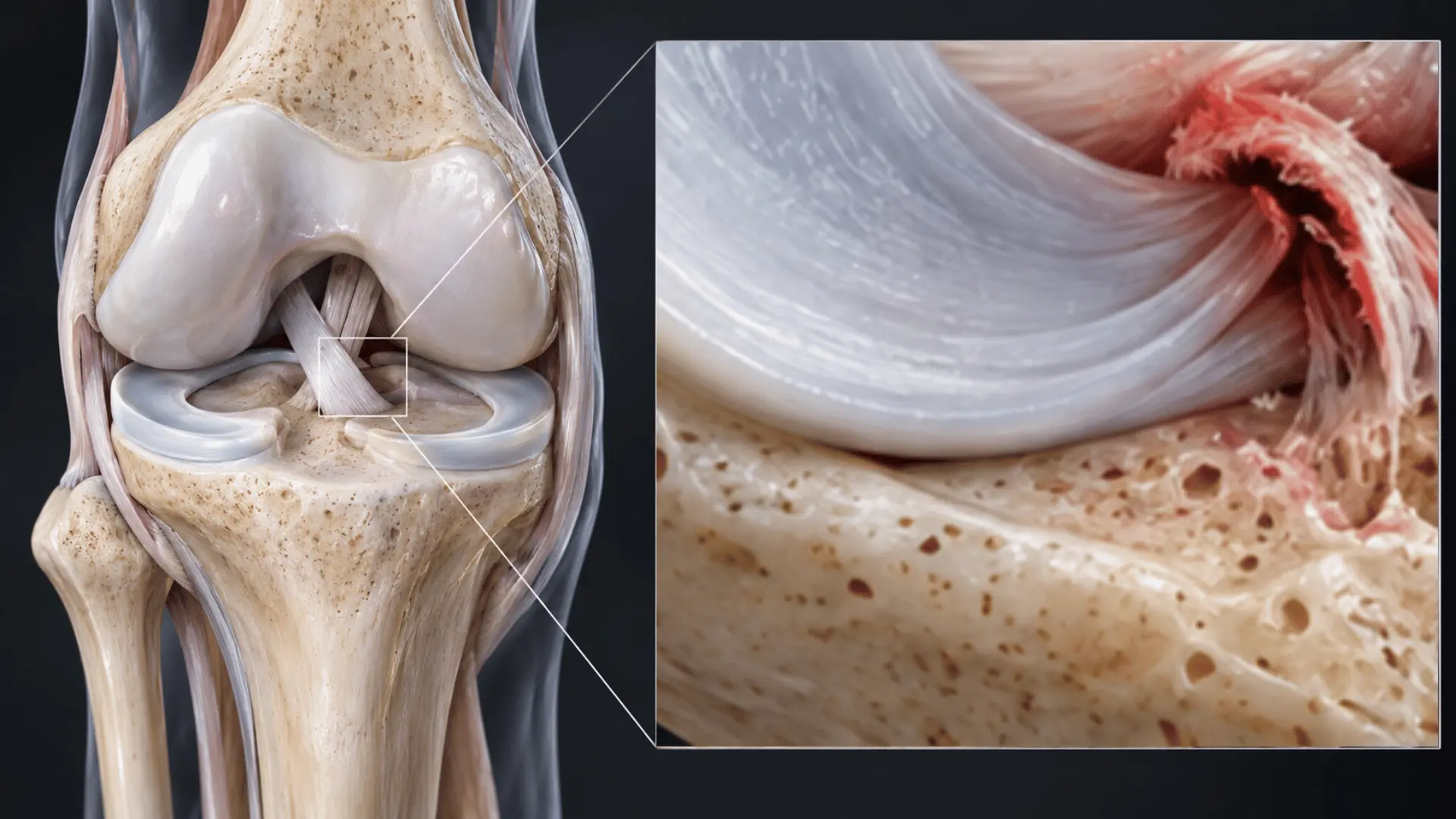
A meniscus root tear is important—but it does not automatically mean surgery.
The meniscal roots anchor the meniscus to the tibia and help it transmit load. When a root is significantly disrupted, the meniscus may lose part of its normal ability to distribute pressure and may move outward from the joint, a finding called meniscal extrusion. The treatment decision therefore needs to consider more than the tear itself: cartilage condition, knee alignment, stability, tear pattern, tissue quality, symptoms, activity demands and patient goals all matter.
“The MRI identifies the structure. The treatment decision depends on the whole knee.”
What exactly is a meniscus root tear?
Each knee contains a medial meniscus on the inner side and a lateral meniscus on the outer side. These fibrocartilage structures help distribute load across the knee, contribute to shock absorption and support normal joint mechanics.
The ends of the menisci are attached to the tibia through the meniscal roots. A root tear involves the attachment itself or a radial tear very close to that attachment. Contemporary reviews commonly describe a complete radial tear within roughly 1 cm of the root attachment as a root-equivalent injury.1
Often a different clinical pattern
Medial posterior root tears may follow a relatively low-energy twist, deep flexion or loading event and may occur alongside degenerative cartilage change.
Frequently linked with ligament injury
Lateral posterior root tears are more commonly associated with traumatic knee injuries, particularly ACL injury, and should be assessed in that context.
The attachment helps control load
A functionally disconnected root can reduce normal hoop-stress transmission and increase contact pressure within the involved compartment.
What can a meniscus root tear feel like?
There is no single symptom that proves a root tear. Some patients remember a specific movement—turning, squatting, rising from a low position or twisting the knee—while others develop pain more gradually.
Joint-line pain, often on the inner side with a medial root tear
Pain with loaded bending, squatting, stairs or prolonged activity
Swelling that appears or increases after activity
Reduced confidence with walking, training or sport
Occasional catching, clicking or a pop around symptom onset
Symptoms that persist despite sensible activity modification
These features are not specific to root tears. Cartilage injury, osteoarthritis, ligament injury, bone-marrow lesions and other meniscal tears can produce overlapping symptoms. Examination and appropriate imaging are therefore important before treatment is selected.
For broader inner-knee pain and meniscus patterns, see Medial Meniscus Tear in Dubai: Inner Knee Pain, MRI Findings & Treatment Options.
What should doctors look for on MRI?
MRI is usually the most useful imaging test when a root injury is suspected because it can assess the meniscus together with cartilage, ligaments, bone marrow and other structures. A 2026 systematic review and meta-analysis of MRI signs found useful diagnostic performance for the cleft/truncated-triangle sign, ghost sign and radial-tear sign.2
| MRI finding | What it may describe | What still needs interpretation |
|---|---|---|
| Root discontinuity / radial defect | A tear involving or very close to the root attachment. | Completeness, tissue quality, chronicity and whether the tear is functionally significant. |
| Ghost sign | Loss or marked reduction of the expected meniscal appearance on selected sagittal images. | Whether the finding is confirmed across imaging planes and matches the overall tear pattern. |
| Cleft / truncated-triangle sign | An abnormal cleft or truncated appearance of the meniscus on coronal imaging. | Location, associated extrusion and the condition of cartilage and subchondral bone. |
| Meniscal extrusion | Outward displacement of the meniscus beyond the tibial margin. | Cause, degree, alignment, cartilage health and whether restoration is realistic. |
| Associated findings | Cartilage loss, bone-marrow change, ACL injury or other structural pathology. | Which finding best explains the patient's symptoms and changes treatment. |
A scan describes structures. A treatment plan should explain function: what happened, what the knee can do now, what is repairable, and what you need to return to.
For a broader explanation of imaging decisions, read MRI for Knee Pain in Dubai: When Do You Really Need It?
What does “meniscal extrusion” mean?
Meniscal extrusion means that part of the meniscus has moved outward beyond its usual position relative to the tibial plateau. It can occur for several reasons and is not exclusive to root tears, but it is commonly associated with medial meniscus root injury and degenerative change.
A threshold of approximately 3 mm has often been used in research to describe substantial extrusion, but measurement methods vary. It should not be treated as a stand-alone surgical threshold.
Do not operate on a measurement.
Extrusion is one piece of the assessment. Symptoms, tear anatomy, cartilage condition, alignment, stability, activity needs and overall repair potential remain essential.
Does a meniscus root tear always need surgery?
No. The diagnosis is important, but the MRI finding alone does not determine treatment.
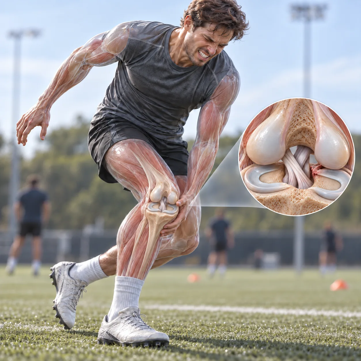
The questions that change the plan
Complete or partial? Acute or longstanding? Repairable tissue or advanced degeneration?
Is the joint surface largely preserved, or is there substantial established osteoarthritis?
Varus or valgus loading can materially change the mechanical environment of the meniscus.
How far has the meniscus displaced, and what does that imply together with the other findings?
Is there associated ACL or other ligament injury that changes the treatment strategy?
Daily walking, work, gym, running and pivoting sport place very different demands on the knee.
Non-operative management
Non-operative care may be reasonable when the overall knee is unlikely to benefit from root repair, surgery is not clinically appropriate, or symptoms and function can be managed satisfactorily without an operation.
- Targeted rehabilitation and progressive strengthening
- Activity and load modification while symptoms settle
- Weight management when clinically relevant
- Medication or bracing in selected cases when appropriate
- Monitoring function and progression rather than treating the MRI alone
Meniscus root repair
Repair may be considered when restoring meniscal function appears biologically and mechanically reasonable—particularly in symptomatic patients with a repairable tear and a joint without advanced irreversible degeneration.
- Repairable root or root-equivalent tear
- Cartilage condition compatible with a preservation strategy
- Alignment and stability evaluated as part of the plan
- Patient able to follow the protected rehabilitation required for healing
- Clear discussion of expected benefits, limitations and alternatives
Repair can be beneficial in appropriately selected knees—but evidence is not a guarantee.
A systematic review of 56 studies and more than 3,000 patients found that root repair produced the greatest numerical improvement in several patient-reported outcomes and appeared to reduce joint-space narrowing compared with partial meniscectomy or non-operative treatment. The authors also emphasised that the literature was heterogeneous and largely Level III–IV evidence.3
This supports a preservation-first discussion when repair is realistic. It does not support a rule that every root tear should be operated on.
For the broader surgical decision, see Meniscus Surgery: Repair, Meniscectomy & Recovery.
Is partial meniscectomy still used for a root tear?
Sometimes, but selectively. Partial meniscectomy removes unstable or irreparable meniscal tissue rather than restoring the root attachment. It may have a role when the tissue cannot realistically be repaired and persistent mechanical symptoms remain.
Because a functionally significant root tear already reduces normal meniscal load transmission, removing additional tissue may further reduce meniscal function. This is why contemporary treatment generally considers meniscal preservation whenever preservation is clinically realistic.3
Does repairing the root stop arthritis?
No procedure can honestly promise that. Root repair aims to restore meniscal function as much as possible and reduce abnormal joint loading. Clinical studies generally report improvements in pain and function after repair in appropriately selected patients, but meniscal extrusion can persist and degenerative changes may continue.
The clinically accurate objective is therefore more measured:
Does the timing of treatment matter?
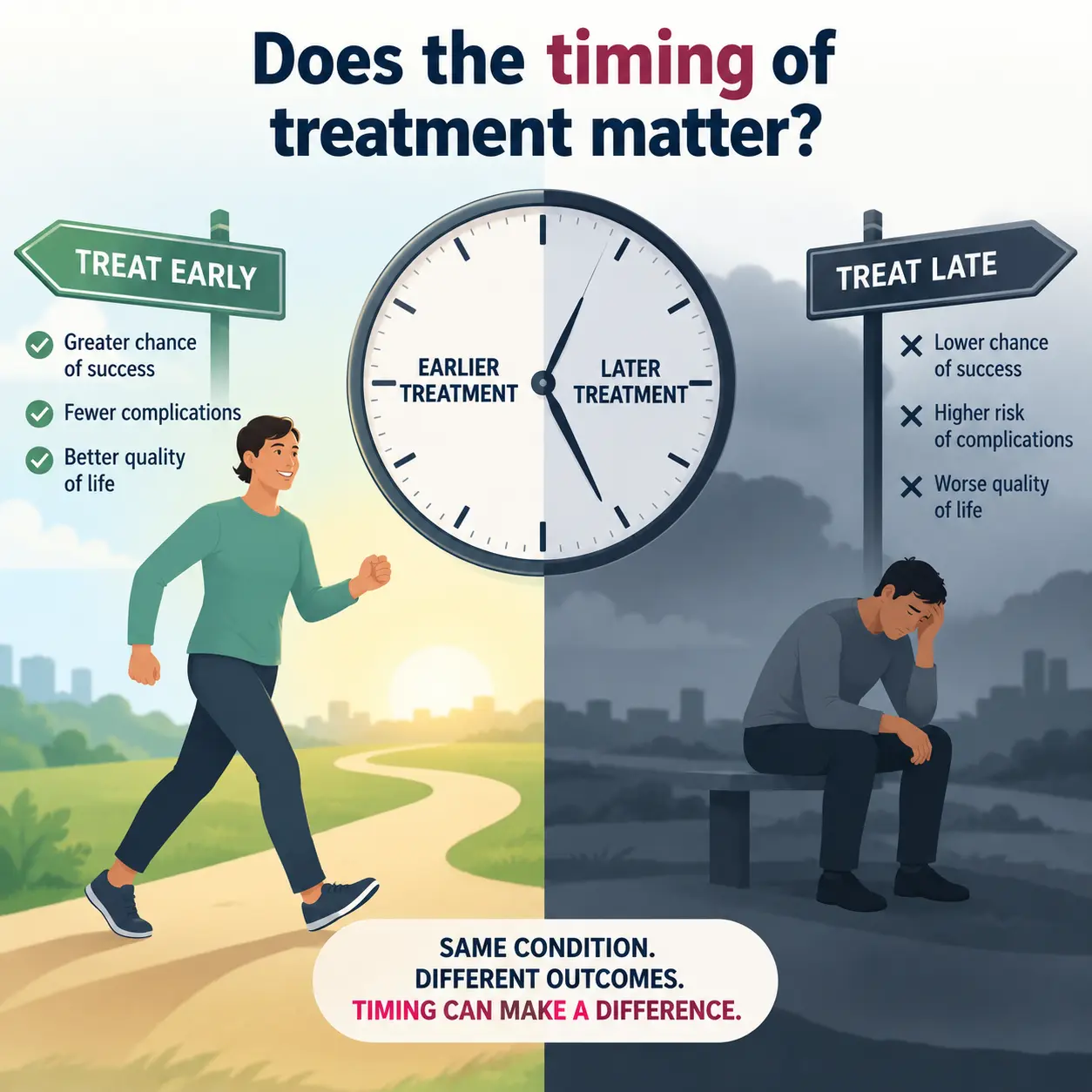
A symptomatic, potentially repairable root tear should not be ignored indefinitely. Recent systematic-review evidence suggests that patients can still improve after delayed medial posterior root repair, while earlier treatment groups have shown greater gains in some patient-reported outcomes. The available studies are heterogeneous, so timing should not be reduced to a rigid cut-off.4
This does not mean that someone who has had symptoms for several months has automatically “missed the window.” It means persistent symptoms and a suspected root tear deserve a proper assessment rather than repeated assumptions that the problem is simply routine knee pain.
Arrange earlier orthopaedic review when:
- Your MRI specifically describes a root or root-equivalent radial tear.
- Swelling repeatedly returns with walking, training or sport.
- The knee has a true mechanical block or cannot fully straighten.
- There is a significant associated ACL or other ligament injury.
- Function is deteriorating rather than improving with a sensible rehabilitation plan.
Seek urgent medical assessment after major trauma if you cannot bear weight, the knee is visibly deformed, swelling or pain is severe and rapidly worsening, the foot is cold or numb, there is fever with a hot red knee, or the knee is truly locked.
Recovery protects healing tissue first—then builds function.
Recovery after root repair is generally more protective than recovery after a straightforward partial meniscectomy because the repaired attachment needs time to heal before progressively greater loads are applied.
The 2024 EU–US Meniscus Rehabilitation Consensus recommends that rehabilitation after meniscus repair progresses using both time and criterion-based milestones. Root, radial and other complex repairs commonly require a more protective early phase than simpler procedures.5
- 1
Protect the repair
Follow procedure-specific restrictions on weight-bearing, brace use and knee flexion while controlling swelling and maintaining safe muscle activation.
- 2
Restore movement and gait
Progress range of motion, quadriceps control and normal walking according to the surgical protocol and tissue response.
- 3
Rebuild capacity
Develop strength, balance and progressively higher functional loads without repeatedly provoking swelling or loss of movement.
- 4
Return with criteria
Running, cutting, jumping and sport should depend on healing time, symptoms, strength, movement quality and task-specific testing—not an arbitrary date alone.
For detailed recovery principles, see Meniscus Surgery Recovery Timeline: Repair vs Trim.
What should a meniscus root tear consultation answer?
A useful consultation should provide more than confirmation that an MRI contains a tear. For patients in Dubai and the UAE, the objective should be to connect the imaging to function and to determine whether preservation, rehabilitation or another pathway is appropriate.
Does this MRI finding explain the symptoms?
Does the tear truly involve the root attachment?
Is the meniscus extruded?
What is the condition of the cartilage and subchondral bone?
What does the knee alignment look like?
Is the knee stable, including the ACL?
Is the tissue realistically repairable?
Can rehabilitation reasonably manage the current problem?
The aim is not to operate because an MRI contains an abnormality. The aim is to understand which treatment is most appropriate for that particular knee.
Meniscus root tear FAQs
These answers are general education. They do not replace examination, review of the original MRI images or an individual treatment plan.
Can a meniscus root tear heal by itself?
A complete root detachment has limited ability to restore its normal anatomical attachment without treatment. However, this does not mean surgery is automatically appropriate. The decision depends on tear pattern, cartilage condition, alignment, symptoms, overall health and realistic repair potential.
Can I walk with a meniscus root tear?
Many patients can walk despite a root tear. The ability to walk does not prove that the meniscus is functioning normally. Loading advice should be individualised, particularly after a recent injury or when swelling increases with activity.
Is a posterior horn tear the same as a root tear?
No. A posterior horn tear can be near the root without disrupting the root attachment. The distance from the attachment, tear orientation and whether the root remains functionally connected all matter.
Does meniscal extrusion mean I need surgery?
No. Extrusion is an important imaging finding, but it should be interpreted with the tear pattern, cartilage condition, alignment, symptoms, stability and treatment goals. It is not a stand-alone indication for an operation.
Am I too old for meniscus root repair?
There is no single age that answers this question. Biological joint condition is often more useful than age alone. Cartilage status, osteoarthritis severity, tissue quality, alignment, medical health, activity requirements and ability to complete rehabilitation should all be considered.
Can root repair prevent knee replacement?
It would be inappropriate to promise that. Repair aims to restore meniscal function and improve the mechanical environment of the knee when possible. Some comparative studies report less progression to arthroplasty after repair than after meniscectomy in selected cohorts, but individual outcomes vary and the evidence is largely observational.
Do I need to bring the MRI images or only the report?
Bring both whenever possible. The written report is useful, but direct review of the original images can clarify tear location, extrusion, cartilage condition, associated ligament injury and other findings that affect treatment.
Patient-information references
- Meniscus root tears: state of the art. International Orthopaedics. 2024.
- Evaluating the diagnostic performance of MRI-based signs for identification of meniscus posterior root tears: a systematic review and meta-analysis. Journal of Orthopaedic Surgery and Research. 2026.
- Root Repair Has Superior Radiological and Clinical Outcomes Than Partial Meniscectomy and Nonoperative Treatment in the Management of Meniscus Root Tears: A Systematic Review. Arthroscopy. 2025.
- Surgical Timing and Outcomes in Medial Meniscus Posterior Root Tear Repair: A Systematic Review. The American Journal of Sports Medicine. 2026.
- The Formal EU-US Meniscus Rehabilitation 2024 Consensus: Part I—Rehabilitation Management After Meniscus Surgery. Orthopaedic Journal of Sports Medicine. 2025.
External references are provided for transparency and patient education. Evidence quality varies between questions, and published studies cannot replace an individual clinical assessment.
A meniscus root tear deserves an explanation—not an automatic procedure.
Dr. Tomislav Cerovecki assesses meniscus injuries, MRI findings, knee stability, cartilage condition and return-to-activity goals at Tadawi Specialty Hospital in Al Garhoud, Dubai. The plan may involve rehabilitation, monitoring, further imaging review or root repair when clinically indicated.
- Doctor
- Dr. Tomislav Cerovecki
- Clinic
- Tadawi Specialty Hospital
- Location
- Al Garhoud, Dubai, UAE
- Clinical focus
- Sports injuries, knee surgery, arthroscopy & joint preservation